The importance of Radiology for immediate diagnosis of Corona virus, COVID-19.
Reference; https://healthcare-in-europe.com/en/news/imaging-the-coronavirus-disease-covid-19.html#
Imaging the coronavirus disease COVID-19
Chest X-ray is the first imaging method to diagnose COVID-19 coronavirus infection in Spain, according to Milagros Martí de Gracia, Vice President of the Spanish Society of Radiology (SERAM) and head of the emergency radiology unit at La Paz Hospital in Madrid, one of the hot spots for viral re-production of COVID-19.
How do these measures affect your department’s workflow?
Milagros Martí de Gracia is Vice President of the Spanish Society of Radiology (SERAM), President of the Spanish Society of Emergency Radiology (SERAU) and head of the department of emergency radiology at La Paz Hospital in Madrid
MMG: ‘The request for chest radiographs has grown exponentially and proportionally with the number of patients visiting the emergency department. A chest X-ray is performed in suspected or confirmed patients through specific circuits.
What is the protocol?
MMG: ‘Patients with respiratory symptoms must remain isolated and wear a mask. If clinical suspicion persists after the examination, a sample of nasopharyngeal exudate is taken to test reverse-transcription polymerase chain reaction (RT-PCR).
Then, we perform a chest X-ray. Getting the results of the PCR test may take several hours. The chest X-ray is a discriminating element; if the clinical situation and the chest X-ray film are normal, patients can go home and wait for the results of the etiological test.
But if the film shows pathological findings, patients are admitted to the hospital for observation.
How important is radiology in diagnosing COVID-19 infection?
MMG: ‘Radiology is fundamental in this process. The radiologist’s main contribution is to facilitate and expedite as much as possible the exploration, help design specific circuits and provide a fast and accurate report of the radiological findings that should indicate whether or not these are consistent with the COVID-19 coronavirus infection.’

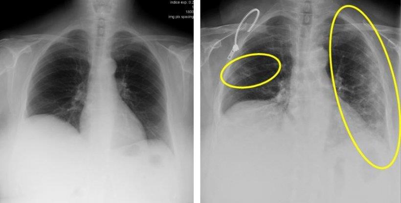
These two X-ray images are from a 72-year-old woman who has a cough and respiratory distress from last year (left) and now. The yellow circle and ovoid indicate the typical subpleural peripheral opacities
What are the typical radiological findings?
MMG: ‘The findings that make us strongly suspect that we are dealing with a COVID-19 infection are the ground glass patterned areas, which, even in the initial stages, affect both lungs, in particular the lower lobes, and especially the posterior segments, with a fundamentally peripheral and subpleural distribution. These findings are present on chest CT in practically 50% of patients in the first two days; For this, in China, CT is being used as a screening or diagnostic method.
‘These lesions progress in the following days until they become more diffuse. If they associate with septal thickening, they will present with a crazy paving pattern. In general, they progress in extension and also towards the consolidation that is done concomitantly with the ground glass pattern, which can present a rounded morphology. It is very rare that it associates lymphadenopathy or capitation or pneumothorax, as the Middle East respiratory syndrome coronavirus (MERS-CoV) did.’
ATX Suggestion:
To supply a vast number of digital X-ray solutions for GP’s / Hospitals and frontline COVID-19 clinics to assist in the immediate diagnosis of COVID19.
The estimated lead time is approximately 7-10 business days however could be expedited substantially given the relevant funding and government assistance.
Prepared: 24/03/20


