atxmarketingATX Medical Solutions

All posts by atxmarketing

ATX Partners With SK Telecom to Introduce AI- Powered Veterinary Aid to the Australian Market

ATX sings partnership agreement with SK Telecom to introduce AI powered veterinary aid to Australia market

On February 13, SK Telecom signed a commercial agreement with ATX Medical Solutions, Australia’s largest medical device distributor, to distribute X Caliber, a veterinary diagnostic assistant, in Australia.

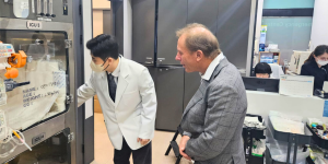
ATX announced on February 13 that it has signed a commercial agreement with SK Telecom (SKT), South Korean wireless telecommunications operator, to distribute X Caliber, Korea’s first AI-based veterinary X-ray image diagnosis assistance service, across Australia.

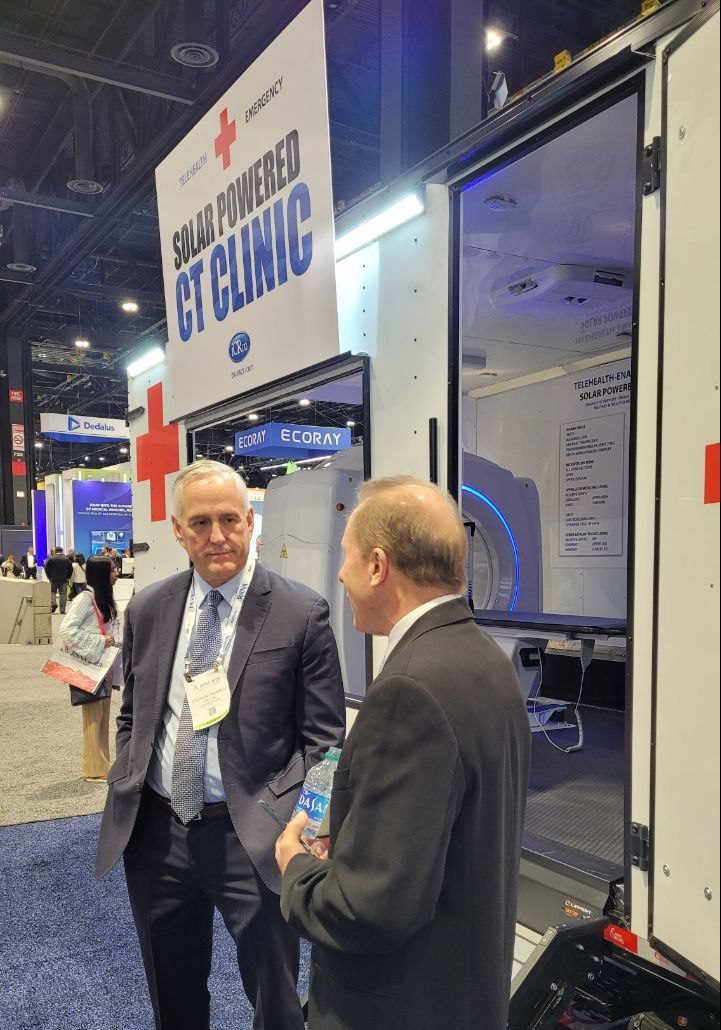
This technology aim to transform veterinary care across Australia, offering rapid, detailed and more effective diagnostics and tailored treatment plans for veterinary practices nationwide.

‘X Caliber’ is a web-based service that uses AI to analyse X-ray images of small animals taken by a veterinarian and uploaded to the AI platform ‘X Caliber Vet AI’, and delivers the analysis results back to the veterinarian within 30 seconds.

Since X Caliber uses cloud to store and retrieve data, there is no need to install a separate server within the hospital. In addition, as a web-based service, X Caliber can be easily upgraded and managed. Veterinarians can examine the results of the AI-based image diagnosis on their mobile devices or PCs anywhere, anytime.

ATX will integrate X Caliber into its cloud software, ITX PACS, a cloud based storage facility that allows to access, store, manipulate and share X-ray data with other computers on the network.

ATX reduces the costs and risks involved with storing these studies and offers a diverse range of tools that have been developed to improve diagnosis and offer effective measurement options for veterinarians to use. Learn more about the ITX Pacs, visit our website: https://atxvet.com.au/services/itx-pacs/

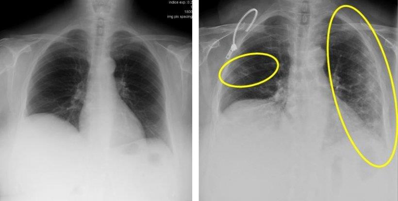
The disease detection rate of the SK Telecom X Caliber stands at 84-86%.* It also showed 97% accuracy in measuring VHS (Vertebral Heart Scale).

  • 84% for detection of 10 different abnormal patterns from chest X-ray images of dogs
  • 86% for detection of 7 different abnormalities from musculoskeletal X-ray images of dogs


The agreement comes less than 100 days after the two companies signed a strategic partnership to utilise X Caliber in November last year.


In the realm of medical imaging, the pursuit of accuracy and reliability is paramount. The choice between large format stitched images and non-stitched ones can play a pivotal role in influencing treatment decisions. In particular, for studies involving the spine and leg length, the techniques used for presenting a contiguous image can significantly impact the diagnostic process. Here are some insights into key techniques for presenting contiguous images. 


Methods to achieve full spine imaging

Full Spine – single TFT panel

The Longtail DR is the only DR system capable of capturing full spine and leg length images in one exposure, without stitching, thanks to its 108cm active area TFT, ensuring complete anatomical coverage and the highest integrity of image.


Multiple Exposures to a Sectional Panel

Normally, 3 to 5 overlapping images are taken to cover the entire anatomy, with manual or automatic stitching. This not only increases patient dose but also introduces the risk of movement and errors in alignment during stitching.


Single Exposure to Multiple Panels

Tipically, 2 or 3 panels are overlapped to capture entire anatomy. Overlap artifacts are eliminated through digital subtraction and images stitched automatically by the software, compromising image integrity.


Single Exposure to Multiple TFT Sheets

Butting up glass TFT sheets may create a seemingly single DR for large images. However, this compromises image integrity, introducing a 4-pixel gap at the glass TFT butt joint. Pixel averaging fills these gaps, leading to a loss of diagnostic information.


How to distinguish high from low integrity large format DR panels

  • System specifications

Check specifications; the format may not always be explicitly stated.


  • Panel Thickness

Is it more than 15mm thick? If so, it is not a single TFT.


  • Exposure to Image Time

Prolonged exposure times (>1-2 seconds) may indicate compromised image integrity.


  • Collimation Capability

Is the panel’s collimation possible anywhere, or is there a specific area for collimated images?


In the pursuit of accurate medical imaging, understanding the nuances of image stitching techniques and their impact on integrity is crucial. By being vigilant about panel specifications, thickness, exposure times, and collimation capabilities, healthcare professionals can make informed decisions to ensure the highest standards of diagnostic accuracy.

ATX Explores New Technologies At Radiological Society of North America Event


Attendance at this event allowed our team to gain insights into advancements by engaging with some of our key radiology equipment partners. These companies showcased their technological innovations, offering a direct glimpse into the future of medical imaging.


Connecting with Our Providers:

Our experience at RSNA Ai enabled us to establish connections with some of our essential suppliers in the industry. We got to engage with CHISON, our recently acquired ultrasound systems provider. They have introduced its latest equipment to ATX, catering to various clinical practice needs, whether it be a cart-based ultrasound, a portable system for on-the-go scanning, or a handheld device for ultimate convenience.

We also had the opportunity to meet up with other providers, including PZ Medical, iRay Technology, CNMC, SYFM, Shielding International, and DEL Medical, among others. Each of these companies showcased their latest innovations during the event.


ATX and DEL Medical Longtail
ATX and Chison ultrasounds systems
ATX and CPI
ATX and PZ Medical
Atx and Shielding international
ATX ans CNMC
ATX and CT Clinic


The AI Showcase: A Hub for Innovation

The RSNA Ai Showcase stands out as the premier space for attendees to connect with companies at the forefront of radiology AI software and products. It serves as a vibrant marketplace where industry leaders and innovators gather to share insights and explore possibilities.

The RSNA Showcase emerged as the focal point for attendees to connect with companies at the forefront of radiology AI software and products. This showcase facilitated interactions between industry leaders and innovators, providing a platform to share insights and explore the endless possibilities within the ecosystem of AI and radiology. We look forward to the ongoing evolution of technology to provide our customers with the best patient care.

ATX team visits Vieworks and NMI offices in South Korea


ATX technical support team in the offices of Vieworks in South Korea


Our technical support team recently embarked on an exciting journey to South Korea, where they had the opportunity to visit the offices of Vieworks and NMI. Both of these companies are renowned manufacturers and industry leaders in the field of X-ray and medical imaging devices. They are our esteemed providers of radiography acquisition systems.

Vieworks and NMI shared their expertise, providing training to our team on X-ray acquisition systems, along with insights into the software components of this equipment. This was a chance for our team to gain a deeper understanding of their X-ray acquisition systems and various radiography solutions available in the market.

We’re excited to implement this newfound expertise to provide our customers with the best quality machines and service.


Strengthened Partnerships

Beyond the technical aspects, this visit strengthened the partnership between our companies. Sharing insights, experiences, and best practices, ensuring that we can continue to provide top-notch solutions and support to our customers across Australia and America.

We look forward to the continued growth of this partnership and the incredible innovations that will result from our collaboration.

The KN95, N95 or P2 Face Masks is perfect for dust protection, influenza protection and general cleaning. This masks offers superior particle filtration over P1, 3 Ply and Surgical Face Masks.

Our KN95 Face Masks are a great solution to keeping you, your business or family safe from pollution, dust, most vapors and influenza. Designed to be worn on the face, the metal clip allows you to adjust the size to your nose gently squeezing the clip whilst the mask is on your face.

The ‘N95’, ‘KN95’ or ‘P2’ rating means that when subjected to careful testing, the respirator blocks at least 95 percent of very small (0.3 micron) test particles. When fitted correctly, the filtration capabilities of KN95 respirators exceed those of surgical or other face masks.

Who should use a KN95/ KN95 Respirator/ Face Mask (Same style as P2 Face Masks)

Disposable KN95, N95 or P2 face masks, when worn correctly will filter out fine dust, water/ influenza particles from the air. Anyone who is working with concrete, underground, around sick persons or looking to reduce exposure to pollution as well as influenza may benefit from wearing a face mask. They reduce exposure to bush fire smoke, concrete dust, saw dust and associated health effects.


Putting on and checking the mask

How to properly wear a KN95 mask or P2 respirator

It is imperative the following checks are completed when still in a safe environment or before entering an area of contamination. Always ensure your P2 respirator/ mask is free from tears or defects every time your mask is used. Ensure when fitting that the KN95 or N95 mask is sealed over the bridge of the nose and around the mouth. Ensure no facial hair, piercings, jewelry or skin conditions are creating gaps.

Always follow manufactures instructions and ensure correct level of protection is chosen for the environment you are entering.

Follow the steps below to correctly fit the KN95 face mask.

  1. Remove any accessories on your head, including glasses, caps or head phones.
  2. Ensuring the metal/ plastic nose clip is over your nose, place the mask over your mouth and nose.
  3. Place the rubber strap or tie the cord around the back of your head of ears (depending on model). The masks should hold itself and should not fall off when you bend over.
  4. While firmly holding the mask over your mouth and nose, pinch/ squeeze the metal/ plastic nose clip to ensure a tight fit over your nose. You want to limit the amount of air which can come through this area.
  5. Check the mask is still sealed around your mouth and chin.
  6. Check you can breath gently inhaling and exhaling. If you notice any air leaks, ensure you adjust the mask and check again. Different styles of masks may have different shapes or leaks if not worn correctly. The mask can take a while to get used to, ensure you maintain a normal and steady breath pattern.

Buy KN95 face masks here

 The importance of Radiology for immediate diagnosis of Corona virus, COVID-19. 

Reference; https://healthcare-in-europe.com/en/news/imaging-the-coronavirus-disease-covid-19.html# 

Imaging the coronavirus disease COVID-19

Chest X-ray is the first imaging method to diagnose COVID-19 coronavirus infection in Spain, according to Milagros Martí de Gracia, Vice President of the Spanish Society of Radiology (SERAM) and head of the emergency radiology unit at La Paz Hospital in Madrid, one of the hot spots for viral re-production of COVID-19.

How do these measures affect your department’s workflow?

Milagros Martí de Gracia is Vice President of the Spanish Society of Radiology (SERAM), President of the Spanish Society of Emergency Radiology (SERAU) and head of the department of emergency radiology at La Paz Hospital in Madrid

MMG: ‘The request for chest radiographs has grown exponentially and proportionally with the number of patients visiting the emergency department. A chest X-ray is performed in suspected or confirmed patients through specific circuits.

What is the protocol?

MMG: ‘Patients with respiratory symptoms must remain isolated and wear a mask. If clinical suspicion persists after the examination, a sample of nasopharyngeal exudate is taken to test reverse-transcription polymerase chain reaction (RT-PCR).

Then, we perform a chest X-ray. Getting the results of the PCR test may take several hours. The chest X-ray is a discriminating element; if the clinical situation and the chest X-ray film are normal, patients can go home and wait for the results of the etiological test.

But if the film shows pathological findings, patients are admitted to the hospital for observation.

How important is radiology in diagnosing COVID-19 infection?

MMG: ‘Radiology is fundamental in this process. The radiologist’s main contribution is to facilitate and expedite as much as possible the exploration, help design specific circuits and provide a fast and accurate report of the radiological findings that should indicate whether or not these are consistent with the COVID-19 coronavirus infection.’

thorax xray images

These two X-ray images are from a 72-year-old woman who has a cough and respiratory distress from last year (left) and now. The yellow circle and ovoid indicate the typical subpleural peripheral opacities

What are the typical radiological findings?

MMG: ‘The findings that make us strongly suspect that we are dealing with a COVID-19 infection are the ground glass patterned areas, which, even in the initial stages, affect both lungs, in particular the lower lobes, and especially the posterior segments, with a fundamentally peripheral and subpleural distribution. These findings are present on chest CT in practically 50% of patients in the first two days; For this, in China, CT is being used as a screening or diagnostic method.

‘These lesions progress in the following days until they become more diffuse. If they associate with septal thickening, they will present with a crazy paving pattern. In general, they progress in extension and also towards the consolidation that is done concomitantly with the ground glass pattern, which can present a rounded morphology. It is very rare that it associates lymphadenopathy or capitation or pneumothorax, as the Middle East respiratory syndrome coronavirus (MERS-CoV) did.’

ATX Suggestion:

To supply a vast number of digital X-ray solutions for GP’s / Hospitals and frontline COVID-19 clinics to assist in the immediate diagnosis of COVID19.

The estimated lead time is approximately 7-10 business days however could be expedited substantially given the relevant funding and government assistance.

Prepared: 24/03/20 

Join our mailing list

and receive special promotions.

Your email is safe with us. We do not share or sell the list to anyone.

  TOP